The binocular indirect ophthalmoscope or indirect ophthalmoscope is an optical instrument worn on the examiners head and sometimes attached to spectacles that is used to inspect the fundus or back of the eye. Abstract Binocular indirect ophthalmoscopy may be performed in the operating room with a fiberoptic surgical headlight and condensing lens eliminating the need for the indirect ophthalmoscope headpiece.
Binocular Indirect Ophthalmoscopy The Beginner S Guide
History of ophthalmoscope Mery in 1704 made first ophthalmoscopic observation of a normal fundus in a drowning cat Cumming and Brucke in 1846 explained the principles of ophthalmoscopy 2.

. In 2003 we introduced the first Keeler wireless indirect ophthalmoscopes available on our All Pupil or Vantage models. It is valuable for diagnosis. How To Perform Indirect Ophthalmoscopy Youtube Binocular Indirect.
It produces an stereo-scopic image with between 2x and 5x magnification. Author M J Garston. Binocular indirect ophthalmoscopy is commonly done with a BIO headset that typically cost between 1000 and 2000.
Wireless gives you the freedom to move from room to room without any constraints no more wires cords or tangles. Put the indirect on and ensure your oculars and light spot are properly centered. Then adjust the pupillary distance and height of the beam so you can see a full beam with each eye.
About Press Copyright Contact us Creators Advertise Developers Terms Privacy Policy Safety How YouTube works Test new features Press Copyright Contact us Creators. Performing Binocular Indirect Ophthalmoscopy is challenging. The binocular indirect ophthalmoscope BIO J Am Optom Assoc.
History of ophthalmoscope Mery in 1704 made first. Preparation Dilate your patient. 411815 Abstract Effective use of diagnostic drugs in primary eye care demands that the optometrist not only be able to better utilize conventional equipment but also utilize new equipment.
For binocular indirect ophthalmoscopy technique doctors use lenses with a magnification of 15 to 30 diopters. In general modern ophthalmoscopes are equipped with LED or XHL lighting. While focusing the light spot at your hand at arms length close one eye at a time to make sure your pupillary distance is properly adjusted and you can see well with each eye.
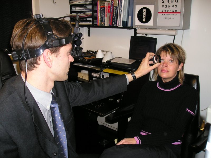
Learning indirect ophthalmoscopy may be the most difficult and stress-provoking exam technique a new resident faces. General procedure for head-band binocular indirect ophthalmoscopy. Teaching video about the correct method of binocular indirect ophthalmoscopy - apt for beginners and residents.
This video will give you a Introduction to using the Binocular indirect ophthalmoscope for fundus examination. See section on mydriasis. The practitioner should be comfortable.
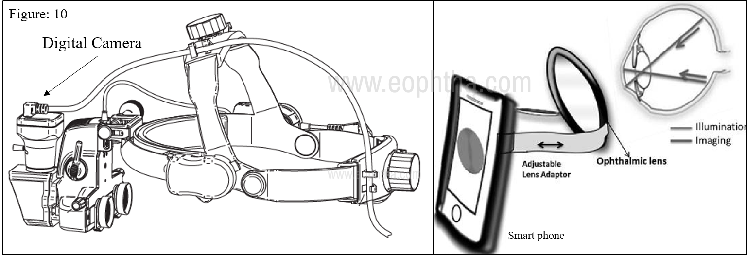
The entire fundus can be seen in 3D up to the far retinal periphery using specifically our OMEGA instruments. This will usually take 20 to 30 minutes following drug instillation. DrShah-Noor Hassan FCPSFRCS Assistant Professor Vitreo-Retina BSMMU.
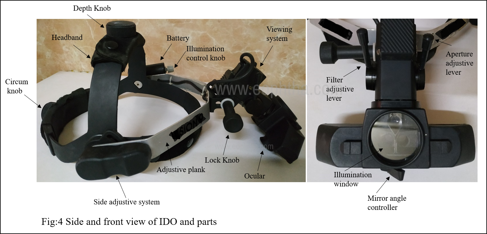
THREE basic principles described by. Binocular Indirect Ophthalmoscopes Indirect ophthalmoscopy is done by ophthalmologists and optometrists to examine the retina as well as the optic nerve vessels and macula lutea. Set the light aperture to the largest spot for a fully dilated patient.
Just a lightweight balanced and comfortable indirect ophthalmoscope. HEINE Hand-held Indirect Ophthalmoscope MONOCULAR. This paper offers a simple and.
Binocular Indirect OPHTHALMOSCOPY 1. History of ophthalmoscope Mery in 1704 made first ophthalmoscopic observation of a normal fundus in a drowning cat Cumming and Brucke in 1846 explained the principles of ophthalmoscopy 2. Adjust the indirect headset First adjust the headband so that the scope is secure on your head.
It will help to darken the room so that minimal lamp intensity can be used. The skill is rightfully challenging indirect ophthalmoscopy proficiency takes tho. We also teach scleral.
It produces an stereoscopic image with between 2x and 5x magnification. Use the smallest aperture for smaller pupils and intraocular gas. Binocular Indirect Ophthalmoscopy.
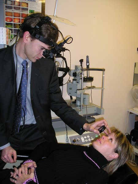
General procedure for slit lamp microscope SLM binocular indirect ophthalmoscopy BIO Preparation Dilate the pupil. The resultant aerial inverted stereo image has enhanced stereopsis but is otherwise identical to that obtained with the headpiece. It will help to darken the room so that minimal lamp intensity can be used.
In this case the field of view decreases. DrShah-Noor Hassan FCPSFRCS Assistant Professor Vitreo-Retina BSMMU 2. The smaller the optical power of the lens is the greater the magnification and working distance turns out to be.
How an indirect ophthalmoscope works The binocular indirect ophthalmoscope or indirect ophthalmoscope is an optical instrument worn on the examiners head and sometimes attached to spectacles that is used to inspect the fundus or back of the eye. Learning indirect ophthalmoscopy may be the most difficult and stress-provoking exam technique a new resident faces. This will usually take 20 to 30 minutes following drug instillation.
See section on mydriasis. This video will give you a Introduction to using the Binocular indirect ophthalmoscope for fundus examination.

3 Examination Of The Fundus Ento Key

Conventional Binocular Indirect Ophthalmoscope Bio A And Portable Download Scientific Diagram
Head Band Indirect Ophthalmoscopy

Inexpensive Binocular Indirect Ophthalmoscopy Technique With 30 Lens Without Bio Headset Youtube
Head Band Indirect Ophthalmoscopy

Binocular Indirect Ophthalmoscopy The Beginner S Guide
How To Use The Indirect Key Tips And Tricks Eyeguru
Binocular Indirect Ophthalmoscopy The Beginner S Guide
0 comments
Post a Comment